Spinal Fusion Instrument Set: Posterior Lumbar Fusion Guide
A complete guide to spinal fusion instruments: pedicle screws, taps, probes, cages and rod fixation for posterior lumbar fusion.
 Made in Sialkot · Since 1980
The patient is prone on a Wilson frame, a degenerative grade-1 spondylolisthesis at L4–L5 confirmed on the lateral fluoro shot. The surgeon has just finished the midline exposure and is reaching for the first instrument that matters: a pedicle awl. From this point until the rods are locked, every step depends on a tray that is laid out correctly and complete. A missing tap or a mismatched screwdriver tip in the middle of a posterior lumbar interbody fusion is not an inconvenience — it stops the case.
A posterior lumbar fusion set is really several instrument families working in sequence: exposure and retraction, pedicle preparation, decompression, interbody work, and finally screw-and-rod fixation. Understanding what each group does — and where the tolerances are unforgiving — is what separates a smooth tray from a scramble.
What a Posterior Spinal Fusion Set Actually Contains
Vendors sell “spinal fusion instruments” as a single SKU, but the working set is modular. Below is the breakdown most spine teams recognise on their count sheet, grouped by the phase of surgery in which each tool is used.
Exposure and Soft-Tissue Retraction
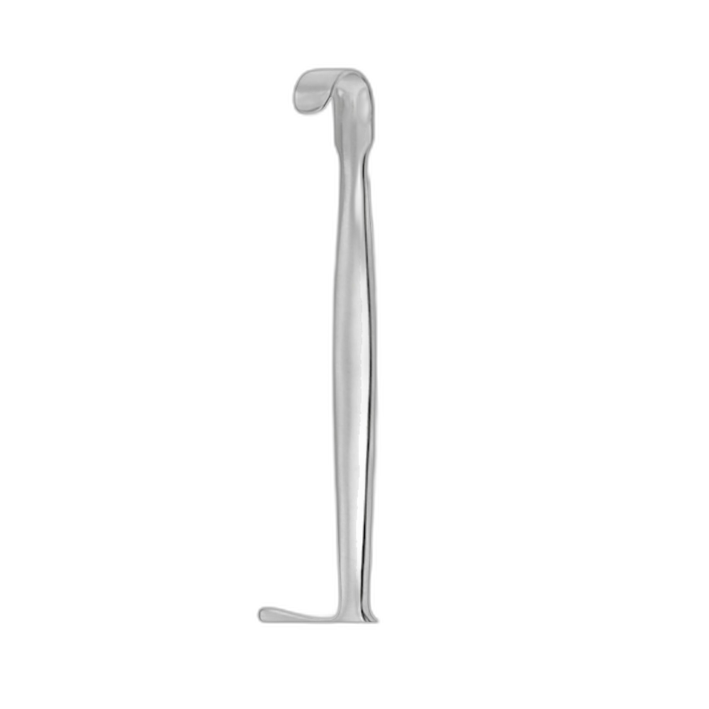
The paraspinal muscles have to be held off the lamina and facet joints for the entire case. A Cobb elevator (usually in 3 widths — 12 mm, 16 mm and 19 mm) strips muscle off bone subperiosteally. Self-retaining retraction is handled by a Taylor retractor anchored into the spinous process, or a table-mounted system for wider levels. Gelpi and Weitlaner retractors hold the superficial layers; for a longer construct, a modular blade system replaces the handheld options.
Deep pituitary rongeurs and a Leksell rongeur clear ligamentum flavum and osteophytes so the pedicle entry points are visible. This is the same rongeur family covered in our guide to orthopedic plating instruments, sized here for the tighter working corridor of the spine.
Pedicle Preparation — The Precision Core
This is where the set earns its keep. Pedicle cannulation follows a fixed sequence, and each instrument has a job the next one depends on:
- Pedicle awl / gearshift probe — creates the starting hole at the junction of the transverse process and superior articular facet.
- Pedicle probe (curved and straight) — advances down the cancellous channel; the surgeon feels for the four bony walls and the floor.
- Ball-tip feeler (sounding probe) — palpates the channel for a breach before any screw goes in. A 2.0 mm ball tip is standard.
- Tap — cuts threads, sized 0.5–1.0 mm under the intended screw diameter.
- Depth gauge — reads screw length off the palpated channel.
Screwdrivers come as a self-retaining shaft plus a range of tips matched to the implant system. A cross-connector and rod persuader complete the reduction hardware.
Decompression Instruments
Kerrison rongeurs (1 mm to 5 mm bites, 40° and 90° up-cutting) remove lamina and undercut the facet. Curettes — straight, angled, and ring — release the disc space and endplates. A nerve root retractor protects the traversing and exiting roots while the interbody space is prepared.
Interbody Cages and Graft Delivery
In a PLIF or TLIF, the disc is removed and the space is filled — this is where fusion actually happens, and it needs its own instrument subset. Disc space preparation uses a sequence of shavers, box cutters, and endplate scrapers to clear cartilage down to bleeding subchondral bone without breaching the endplate. Paddle distractors and trial spacers size the interbody device.
The cage itself — PEEK or titanium, packed with autograft, allograft, or a bone-graft substitute — is delivered on an inserter that holds it square to the trajectory. Getting the trial sizing right matters: an undersized cage subsides into the endplate and loses correction; an oversized one risks endplate fracture or nerve traction on insertion. A funnel and bone tamp deliver graft around the cage to build the fusion bed. In a TLIF the working corridor is a single Kambin’s triangle, so the inserters are angled to sweep the cage across the disc space from one side.
Graft harvesting adds its own instruments if local bone is insufficient — a gouge and mallet for iliac crest, or simply the bone captured from the decompression, morselised in a bone mill. The morselised local bone from the laminectomy is often the best graft available and should never be discarded.
Intraoperative Imaging and Navigation
Screw accuracy is confirmed, not assumed. Traditional fluoroscopy uses a C-arm for AP and lateral shots to check trajectory and depth against the pedicle and endplate. Increasingly, navigation and robotic systems reference a patient tracker to a preoperative or intraoperative CT, and the pedicle instruments carry reflective arrays so the awl and probe positions render live on screen.
Navigation changes the tray. Navigated awls, probes, taps, and drivers are dedicated instruments with mounting geometry for the tracker array — they are not interchangeable with the standard open set, and a spine service running both open and navigated cases has to stock both. The instruments still do the same job; they simply report where they are while doing it.
Pedicle Screw and Rod Specifications
The implant sizing is where procurement teams get tripped up, because a set has to span the anatomy of an entire caseload, not one patient. These are the ranges a lumbar tray is expected to cover:
| Component | Typical Range | Notes |
|---|---|---|
| Polyaxial screw diameter | 5.5 – 8.5 mm | 0.5 mm increments; 6.5/7.5 mm most used in lumbar |
| Screw length | 30 – 60 mm | 5 mm increments; L5/S1 often 45–50 mm |
| Rod diameter | 5.5 mm (6.0 mm cobalt-chrome option) | Titanium alloy standard |
| Set screw torque | Per system (often 8–12 Nm final) | Torque-limiting driver mandatory |
| Tap undersize | 0.5 – 1.0 mm below screw OD | Bone quality dependent |
Implants themselves are titanium alloy (Ti-6Al-4V) for its modulus closer to bone and its MRI compatibility. The reusable instruments — probes, taps, screwdrivers — are martensitic surgical stainless, hardened for edge retention on the tap and toughness on the drivers.
Material and Manufacturing Standards
Reusable spinal instruments live a hard life: repeated autoclave cycles, high torque on the drivers, and impaction loads on the awl. Fizza manufactures these from AISI 420 and 465-series stainless conforming to ISO 7153-1, the standard governing metallic materials for surgical instruments. Cutting components such as taps are through-hardened to 48–52 HRC so the thread cutters stay sharp across dozens of cases; drivers are tempered slightly softer to resist snapping under torque.
Every instrument is passivated to build the chromium-oxide layer that resists the pitting corrosion autoclaving would otherwise cause. This is the same corrosion-resistance discipline behind our ISO 13485 and CE quality system, and it is why a properly finished pedicle probe will outlast the implant systems it serves.
Setting Up the Tray: Count-Sheet Logic
A spinal fusion tray is counted twice — before incision and before closure — because the consequences of a retained instrument in the paraspinal space are severe. Organise the tray in the order of use, not alphabetically: exposure at the top, pedicle preparation in the centre stringer where the scrub tech reaches most often, decompression rongeurs and curettes to one side, and fixation hardware staged last.
Keep taps and screwdriver tips on a dedicated caddy. Mixing a 6.5 mm tap back into a 7.5 mm slot is the single most common cause of a mid-case pause. For teams building a new spine service line, cross-reference this layout with our broader guidance on general bone surgery instruments to avoid duplicate purchasing across trays.
Reprocessing Spinal Instruments
Cannulated probes and taps trap bone debris and marrow. These must be brushed through the lumen with a sized cleaning brush before enzymatic soak — a visual inspection of the channel under magnification is not optional for cannulated tools. Torque drivers should be checked against a calibration jig periodically; a driver that under-reads risks under-seating a set screw, and rod dislodgement is a return-to-theatre event.
Insulated and ratcheted instruments get inspected for cracked coatings and sluggish action every cycle. Anything with a compromised tip goes out of service — a rounded pedicle probe tip loses the tactile feedback the surgeon relies on to feel a medial wall breach.
Frequently Asked Questions
How many instruments are in a standard posterior lumbar fusion set?
A single-level posterior lumbar set typically runs 30–40 reusable instruments plus the implant caddy. Multi-level or deformity constructs add more rod persuaders, reduction towers, and longer screwdrivers, pushing a full deformity set past 60 pieces across two or three trays.
What is the difference between monoaxial and polyaxial pedicle screws?
A monoaxial screw has a fixed head aligned with the shaft, giving maximum corrective control for deformity work. A polyaxial screw has a head that pivots, making rod capture far easier when screw trajectories differ between levels. Most degenerative lumbar fusions use polyaxial screws for that reason.
Why are pedicle instruments cannulated?
Cannulation allows the instruments to pass over a guidewire in percutaneous and navigated cases, and it lets the surgeon confirm channel position with imaging. The trade-off is reprocessing: the lumen must be brushed and inspected under magnification every cycle to prevent retained bone debris.
What steel are reusable spinal instruments made from?
Reusable probes, taps, and drivers are martensitic surgical stainless (AISI 420/465 family) hardened for edge retention and torque strength, conforming to ISO 7153-1. The implants themselves — screws and rods — are titanium alloy for its bone-like modulus and MRI compatibility.
Can one set cover both open and minimally invasive fusion?
Partly. The pedicle preparation and fixation logic is identical, but MIS cases need tubular retractors, guidewires, and extended-tab reduction screws that an open set does not carry. Many hospitals buy the open set first and add an MIS module as case volume grows.
Choosing a Manufacturing Partner
Spinal instruments are unforgiving of poor finishing. A tap that loses its edge, a probe tip that mushrooms under impaction, or a driver that fails its torque calibration all translate directly to time on the table. Fizza Surgical manufactures reusable spinal fusion instruments in Sialkot under an ISO 13485 quality system, CE marked and finished to the same corrosion and hardness standards demanded across our surgical range. For hospitals standardising a spine service line, matched sets with documented material certificates remove the guesswork from procurement.
Need precision surgical instruments?
Configure complete instrument sets with our team — ISO 13485 certified, CE marked, made in Sialkot since 1980.
Related Guides
View all articles →
Where We Serve
Fizza Surgical exports to 50+ countries. Browse our country-specific pages with local regulatory guidance and pricing: