Parotidectomy Instrument Set: Facial Nerve Dissection Guide
Parotidectomy instruments tray by tray: McCabe dissector, fine mosquitoes, bipolar, nerve monitoring and a full set list with sizes.
What does a parotidectomy tray actually need that a general soft-tissue set does not?
Fewer instruments than most people expect — but finer ones, and a handful that cannot be substituted. The operation is defined by a single structure: the facial nerve, running through the gland in a plane that is often only a few millimetres from a tumour capsule. Every instrument choice on the tray follows from the need to find that nerve, follow it and leave it working.
This guide works through the parotidectomy instruments in the order the surgeon uses them, from skin incision to closure, then sets out a complete tray list, how the set changes for superficial, total and extracapsular procedures, and what to inspect on the fine instruments that do the nerve work.
The Short Version: Fine Instruments, a Stimulator and Good Light
Head and neck teams often say that parotid surgery needs no specialised instrumentation, and in one sense that is true. There is no parotid-specific retractor or clamp that the operation cannot be done without. What the procedure demands instead is a particular standard of ordinary instruments:
- Scissors and mosquito forceps with fine, perfectly aligned tips for spreading tissue along the nerve
- A fine-tipped bipolar forceps, because monopolar current near the facial nerve is a risk most surgeons will not take
- A facial nerve monitoring system with a sterile stimulating probe
- Magnification — surgical loupes at 2.5× to 3.5× are the norm, with a microscope used by some surgeons for branch dissection
- A dedicated nerve dissector such as the McCabe pattern
Everything else on the tray is a well-chosen general soft-tissue set. The mistake procurement teams make is not missing an exotic instrument; it is putting heavy general-surgery scissors and worn hemostats in a tray where the operation depends on tip precision.
Opening: Incision and Skin Flap Instruments
Most parotidectomies use a modified Blair (“lazy S”) incision: down in the preauricular crease, around the lobule, and curving forward into a natural neck crease about two fingerbreadths below the mandible. Some surgeons use a facelift-style incision for cosmetic reasons, which puts more demand on flap retraction.
What the opening needs
- Scalpel: No. 3 handle with #15 blades for the skin incision; a #12 blade is useful later for dividing parotid tissue over a known nerve line.
- Skin hooks: double-prong hooks (Gillies or Joseph pattern) to lift the flap edge with minimal crushing. Our skin hooks guide compares the patterns.
- Tissue forceps: Adson toothed forceps, 12 cm, for handling skin edges.
- Flap elevation: Metzenbaum scissors, 14.5 cm, or Stevens tenotomy scissors for a thinner flap raised in the plane just superficial to the parotid fascia.
- Retraction: Senn double-ended retractors and small rake retractors early on; many surgeons then stitch the anterior flap forward rather than holding it.
Two nerves are at risk before the gland is even reached. The great auricular nerve crosses the sternocleidomastoid in the posterior part of the field, and its posterior branch is preserved where oncologically possible. Fine scissors and a gentle retractor protect it far better than blunt finger dissection.
Finding the Facial Nerve Trunk: Landmarks and the Instruments That Reach Them
The antegrade approach — identify the main trunk first, then follow each branch forward — is the standard technique. It relies on separating the gland from the cartilaginous ear canal and the mastoid, then working deep in a small, bloody space. This is where the tray earns its money.
The four landmarks
| Landmark | Relationship to the facial nerve trunk | Instruments used to expose it |
|---|---|---|
| Tragal pointer | Trunk lies roughly 1 cm deep and slightly anterior-inferior to the tip of the cartilage | Freer elevator, fine curved scissors, Senn retractor |
| Tympanomastoid suture | Trunk exits 6–8 mm deep to the point where the suture drops off | Mosquito forceps, bipolar forceps, fine suction |
| Posterior belly of digastric | Trunk lies at about the same depth, just superior to the muscle’s upper border | Small rake retractor, Metzenbaum scissors |
| Styloid process | Trunk passes lateral to its base | Palpation with a fine instrument; rarely exposed deliberately |
Surgeons rarely rely on one landmark alone. The trunk is confirmed where two or three of them converge, then verified with the stimulator.
The dissecting instruments
Fine curved mosquito forceps. Hartmann or Jacobson patterns, 10–12.5 cm, with delicate jaws. The classic nerve dissection move is to slide the closed tips along the nerve in the plane superficial to it, open them to spread the overlying gland, and then divide the tented tissue. Tip alignment is everything: a mosquito whose jaws cross or gape at the tip will tear a nutrient vessel or catch the nerve sheath.
McCabe facial nerve dissector. An angled instrument with smooth, blunt jaws (around 14 cm long) designed to elevate the superficial lobe off the nerve and follow the trunk distally. The smooth jaws spread tissue without cutting it. For related instruments used in nerve work elsewhere, see our nerve hooks and micro dissectors guide.
Fine scissors. Stevens tenotomy or Jameson scissors with blunt tips for dividing the glandular bridge above the nerve once it has been spread. Sharp-tipped iris-style scissors are generally avoided in this step.
Suction. A Frazier suction tube in a small size — 7 or 9 Fr — keeps a dry field without the suction force that pulls on delicate branches. The thumb-port control lets the surgeon drop suction to almost nothing near the nerve.
Retrograde dissection when the trunk is buried
A large tumour sitting over the trunk, a recurrence with scar tissue, or a deep-lobe mass can make the antegrade approach unsafe. The alternative is retrograde dissection: find a peripheral branch and trace it back toward the trunk.
The two common starting points are the marginal mandibular branch, where it crosses the facial vessels below the mandible, and the buccal branch, which runs alongside Stensen’s duct. Retrograde work calls for longer, finer instruments and more magnification, because the branches are thinner and the dissection is longer. Trays for revision cases benefit from an extra fine mosquito and a second McCabe dissector.
Facial Nerve Monitoring Equipment
Intraoperative monitoring does not replace anatomical identification, but in most units it has become routine. It is especially valuable in revision surgery, deep-lobe tumours and cases where inflammation has distorted the tissue planes.
A typical setup includes:
- Recording electrodes: paired needle electrodes placed in facial muscles — commonly orbicularis oculi and orbicularis oris, with frontalis and mentalis added on four-channel systems.
- Stimulating probe: a sterile monopolar or bipolar probe, reusable or single-use depending on the system.
- Stimulation settings: these vary by protocol and system. Many surgeons search with a higher current, then reduce it to confirm individual branches; published studies use settings in the range of roughly 0.5–2 mA. Follow the manufacturer’s guidance and your department’s protocol rather than a number from a tray list.
- Anaesthesia coordination: long-acting neuromuscular blockade suppresses the EMG response, so the anaesthetic plan has to allow for it.
From an instrument-set perspective, the practical point is that a reusable probe belongs in the tray count and needs its own inspection for insulation damage, just like bipolar forceps.
Haemostasis Near the Nerve: Bipolar, Clips and Ties
Bleeding in the parotid bed obscures the nerve plane faster than almost anything else, so haemostasis instruments deserve careful selection.
- Bipolar forceps: fine-tipped, around 0.5–1 mm tips, straight or bayonet, 18–19 cm. Bipolar confines current between the tips, which is why it is preferred near neural tissue. Our comparison of monopolar vs bipolar instruments explains the thermal spread difference.
- Monopolar: useful for the skin flap and posterior dissection away from the nerve, but most surgeons switch it off once the nerve plane is approached.
- Ligating clips: small clips with a matching applicator for vessels such as branches of the retromandibular vein.
- Ties: 3-0 and 4-0 absorbable ties passed with a fine right-angle or mosquito.
- Energy devices: ultrasonic and advanced bipolar devices are used in some centres. Comparative studies suggest they can reduce operating time and drain output, but they add single-use cost and are not a substitute for fine forceps.
Closure, Drains and Frey Syndrome Prevention
Closure gets less attention than nerve dissection, yet it is where two common complications — seroma and gustatory sweating — are either prevented or set up.
Drains
Most surgeons leave a closed suction drain, typically 10 Fr, brought out through a separate stab incision behind the lower end of the wound. The tray needs a trocar or a large curved artery forceps to pull the drain through, a 2-0 or 3-0 non-absorbable suture to secure it, and scissors that will cut drain tubing cleanly without crushing it. Some surgeons skip the drain after small extracapsular resections, so it is sensible to keep drains in the sterile supply rather than packed into every tray.
Reducing the dead space
Frey syndrome — sweating and flushing over the cheek when eating — results from severed parasympathetic fibres regrowing into the sweat glands of the overlying skin. Techniques that place a barrier between the parotid bed and the skin flap, such as a sternocleidomastoid muscle flap or plication of the superficial musculoaponeurotic layer, aim to reduce it. For the instrument set, that means a pair of non-toothed dissecting forceps, 3-0 absorbable sutures on a small curved needle and a needle holder with enough length to work under the raised flap.
Skin closure
The deep layer is closed with 3-0 or 4-0 absorbable sutures and the skin with a 4-0 or 5-0 subcuticular or fine interrupted suture. A tungsten carbide needle holder of 13–15 cm grips these small needles without rotating them, and a fine Adson forceps everts the edges for a neat preauricular scar — which patients notice far more than surgeons expect.
Complete Parotidectomy Instruments Tray List
The table below reflects a practical superficial parotidectomy set for an adult patient. Quantities are a sensible starting point rather than a universal standard; individual surgeons will adjust.
| Instrument | Size / pattern | Qty | Role |
|---|---|---|---|
| Scalpel handle | No. 3, with #15 and #12 blades | 2 | Skin incision; dividing gland over the nerve line |
| Adson tissue forceps | 12 cm, 1×2 teeth | 2 | Skin edge handling |
| DeBakey tissue forceps | 15 cm, 1.5 mm atraumatic | 2 | Gland and vessel handling |
| Double skin hooks | Gillies or Joseph, 16 cm | 2 | Flap elevation |
| Metzenbaum scissors | 14.5 cm, curved | 1 | Flap and posterior dissection |
| Stevens tenotomy scissors | 11.5 cm, curved, blunt | 2 | Dividing gland above the nerve |
| Mayo scissors | 15 cm, straight | 1 | Sutures |
| Mosquito forceps | Hartmann/Jacobson, 10–12.5 cm, curved | 8 | Spreading along the nerve; haemostasis |
| Crile artery forceps | 14 cm, curved | 4 | Larger vessels, retraction of gland tissue |
| McCabe facial nerve dissector | ~14 cm, smooth angled jaws | 1 | Elevating gland off trunk and branches |
| Freer elevator | Double-ended, 19 cm | 1 | Separating gland from ear canal cartilage |
| Senn retractors | Double-ended, 16 cm | 2 | Superficial retraction |
| Small rake retractors | 3- or 4-prong, blunt | 2 | Flap and SCM retraction |
| Frazier suction tubes | 7 Fr and 9 Fr | 2 | Fine suction near the nerve |
| Bipolar forceps | 18–19 cm, 0.5–1 mm tips | 1 | Haemostasis in the nerve plane |
| Ligating clip applicator | Small | 1 | Vessel control |
| Needle holder | Crile-Wood or Webster, 13–15 cm, TC jaws | 2 | Subcuticular and skin closure |
| Nerve stimulator probe | System-specific, reusable or single-use | 1 | Nerve identification and confirmation |
| Towel clips | Backhaus, 11 cm | 4 | Draping |
Materials: scissors in AISI 420 hardened stainless steel for edge retention; forceps and retractors in AISI 410 or 420; tungsten carbide inserts for needle holders handling fine 4-0 and 5-0 sutures. Surface finish should be satin to reduce glare under loupes and a headlight.
How the Set Changes: Superficial, Total and Extracapsular
Not every parotid operation uses the same tray, and it pays to plan add-on packs rather than one oversized set.
Extracapsular dissection
For small, mobile benign tumours in the superficial lobe, some surgeons remove the tumour with a margin of normal tissue without formally dissecting the nerve trunk. The set can be trimmed: fewer mosquitoes, no need for the full retraction complement, but the stimulator stays. Monitoring matters more, not less, when the nerve is not being visually traced.
Superficial parotidectomy
The tray above covers this — the most common procedure for benign lesions such as pleomorphic adenoma and Warthin tumour.
Total and deep-lobe parotidectomy
Removing tissue deep to the nerve means working beneath retracted branches. The add-on pack typically includes vessel loops for gentle branch retraction, additional fine mosquitoes, a small self-retaining retractor such as a Weitlaner, and longer bipolar forceps. Deep-lobe tumours extending into the parapharyngeal space may require exposure that borrows from a neck dissection tray.
Combined with neck dissection
Malignant parotid tumours frequently need a neck dissection in the same sitting. In that case the parotid set is opened alongside a head and neck major tray rather than duplicating long retractors and heavy clamps. Our neck dissection instrument set guide lists that tray, and the thyroidectomy set shows how a similar fine-dissection philosophy applies to the recurrent laryngeal nerve.
Buying and Maintaining the Nerve-Dissection Instruments
Among parotidectomy instruments, a small number carry most of the clinical risk. Those are the ones to specify tightly and inspect every cycle.
Mosquito forceps
- Jaws should meet along their full length with tips aligned — no crossover when closed under light pressure.
- Serrations should be fine and complete to the tip; worn tips slip on tissue and surgeons compensate with force.
- The box joint should move smoothly without lateral play.
McCabe dissector and fine scissors
- Check the smooth jaws for burrs or scratches under magnification — a raised burr on a nerve dissector defeats its purpose.
- Test tenotomy scissors on appropriate test material at incoming inspection and after each service. Blunt scissors crush the glandular bridge rather than dividing it, which increases traction on the nerve.
Bipolar forceps and the stimulator probe
- Inspect insulation for cracks, particularly near the tips and at the cable connector.
- Check tip alignment; misaligned bipolar tips spread heat unevenly and stick to tissue.
- Follow the system manufacturer’s reprocessing limits for reusable probes.
For tenders, list each fine instrument separately with its pattern, length, tip form and steel grade rather than accepting a generic “ENT soft-tissue set” line item. Distributors supplying head and neck units should also ask for batch traceability on each instrument.
Fizza Surgical manufactures the soft-tissue and ENT instruments in this tray in Sialkot under an ISO 13485 quality management system with CE marking. Our certifications page and surgical instruments catalogue are good starting points for building a parotidectomy instruments specification.
Frequently Asked Questions
What instruments are used in a parotidectomy?
A fine soft-tissue set: #15 blades, skin hooks, Adson and DeBakey forceps, Metzenbaum and Stevens tenotomy scissors, curved mosquito forceps, a McCabe facial nerve dissector, Senn and rake retractors, fine Frazier suction, bipolar forceps, and a facial nerve monitoring system with a sterile stimulating probe.
Is facial nerve monitoring mandatory?
It is not universally mandatory, and it does not replace anatomical identification of the nerve. However, it is now routine in many units and is particularly valuable in revision surgery, deep-lobe tumours and cases with distorted anatomy.
Why is bipolar preferred over monopolar near the facial nerve?
Bipolar current passes only between the two tips of the forceps, which limits lateral thermal spread. Monopolar current travels through the patient to a return electrode and carries a greater risk of heat injury to nearby nerve branches.
What is a McCabe dissector?
It is an angled facial nerve dissector with smooth, blunt jaws, about 14 cm long, used to elevate parotid tissue off the facial nerve trunk and its branches by spreading rather than cutting.
Does a total parotidectomy need a different tray?
It uses the same core set plus an add-on pack: vessel loops for branch retraction, extra fine mosquito forceps, a small self-retaining retractor and longer bipolar forceps. If a neck dissection is performed at the same time, a head and neck major tray is opened alongside.
Need precision surgical instruments?
Configure complete instrument sets with our team — ISO 13485 certified, CE marked, made in Sialkot since 1980.
Related Guides
View all articles →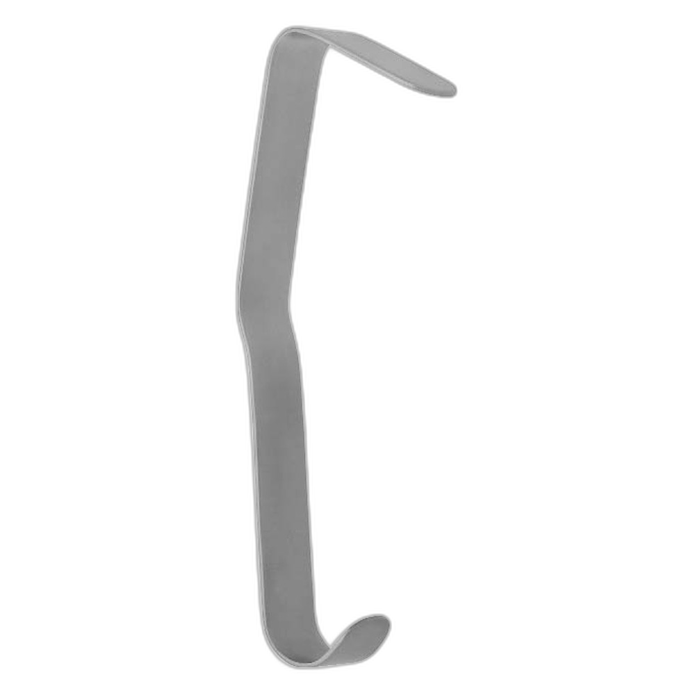


Where We Serve
Fizza Surgical exports to 50+ countries. Browse our country-specific pages with local regulatory guidance and pricing: