Sequestrum Forceps and Bone Nibblers: Osteomyelitis Instruments
Sequestrum forceps and bone nibblers for sequestrectomy: patterns, sizes, cavity saucerisation and reprocessing.
Before antibiotics, chronic osteomyelitis was a surgical disease treated almost entirely with steel. The dead bone had to come out, the cavity had to be laid open, and the instruments that did that work — long serrated grasping forceps and nibbling punches — were among the most-used items in any nineteenth-century orthopaedic set.
Antibiotics changed the incidence, not the principle. A sequestrum has no blood supply, so no systemic drug reaches it. It is still removed mechanically, and the instruments are recognisably the same.
What a Sequestrum Is, and Why It Must Be Removed
In chronic osteomyelitis, infection strips periosteum from cortex and thromboses the intracortical vessels. The devascularised segment dies but remains in place, walled off inside a shell of reactive new bone — the involucrum — with sinus tracts draining through it.
That dead fragment is the sequestrum. It is avascular, so antibiotic penetration is effectively zero, and its surface carries a biofilm that host defences cannot clear. As long as it stays, the infection recurs. Sequestrectomy — surgical removal of the sequestrum together with the surrounding infected granulation tissue — is the definitive step.
The operation has a predictable instrument sequence: expose, window the involucrum, grasp and extract the dead bone, then debride and saucerise the cavity.
Sequestrum Forceps: Design and Patterns
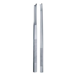
Sequestrum forceps are long grasping forceps with straight or side-angled jaws, serrated on the inner faces, built to reach into a bone cavity and hold a hard, irregular, often slippery fragment while it is worked free.
Three design features distinguish them from a general-purpose artery or tissue forceps:
- Length. Typically 18–23 cm, because the working end has to pass through a cortical window and reach the depth of a medullary cavity.
- Jaw serration. Coarse cross-serrations, not fine longitudinal grooves. The grip has to hold bone under rotational load, not soft tissue under gentle traction.
- Jaw robustness. Heavier stock than a soft-tissue forceps, since extraction often means levering and twisting rather than a clean pull.
The best-known pattern is the Van Buren, supplied straight or angled to the side. The side-angled version is the more useful of the two in a deep cavity — it keeps the surgeon’s hand and the shaft out of the line of sight while the jaw works at the base of the defect.
| Pattern | Length | Jaw | Best use |
|---|---|---|---|
| Van Buren, straight | 20–23 cm | Serrated, straight | Direct-line access through a wide cortical window |
| Van Buren, angled to side | 23 cm | Serrated, side-angled | Deep medullary cavity, restricted window |
| Standard sequestrum forceps | 18–20 cm | Serrated, straight | General sequestrectomy, small tubular bones |
| Fergusson lion-jaw | 18 cm | Toothed | Grasping and stabilising larger bone segments |
Steel is typically AISI 420 hardened to 46–50 HRC through the jaw. Softer jaws spread under load and stop meeting at the tip; harder jaws risk chipping a serration when they close on cortical bone with nothing between them.
Bone Nibblers: Opening and Shaping the Cavity
A bone nibbler — the term is used interchangeably with rongeur in most theatres — is a punch-cutting forceps that removes bone in small bites rather than in a single cut.
In sequestrectomy it does two jobs:
Windowing the involucrum. The reactive shell over a sequestrum is often thick and dense. The nibbler enlarges an initial drill hole or osteotome cut, biting the margin back until the window is wide enough to admit the sequestrum forceps and let the fragment out. That last point governs the size: the window must be larger than the widest dimension of the dead bone, not merely larger than the instrument.
Saucerisation. After extraction, the cavity walls are cut back to a shallow, saucer-shaped defect with no undercut edges and no dead space where fluid can collect. The nibbler shapes those margins, and rounding sharp edges also protects the soft-tissue closure that goes over them.
Patterns commonly used are Luer, Stille-Luer, Beyer, and Leksell for the heavier cortical work, and Kerrison-type punches for fine margin trimming in narrow access. Single-action jaws suit thin bone; double-action gives more mechanical advantage on dense involucrum with less hand fatigue.
The Rest of the Sequestrectomy Set
Neither forceps nor nibbler works alone. A functional set carries:
- Periosteal elevators — Bristow, Cobb, or Freer, to strip periosteum and define the involucrum
- Osteotomes and gouges with a mallet, for the initial cortical window
- Bone curettes in graduated sizes — Volkmann or Spratt patterns — to scrape granulation tissue and residual necrotic bone from the cavity walls
- Sinus forceps and a probe director to trace and excise the draining tracts
- Suction with a large-bore tip, plus pulsed lavage for debris clearance
- Specimen containers — multiple, since separate tissue samples for culture and histology from different cavity zones materially change antibiotic selection
That last item is not an afterthought. Sequestrectomy is one of the procedures where instrument set design should explicitly account for specimen handling, because a single pooled swab from a chronically infected cavity is close to useless microbiologically.
Instrument Care in an Infected Field
Sequestrectomy instruments come off the field carrying necrotic bone, biofilm, and purulent debris packed into serrations and jaw hinges. Reprocessing failures here are consequential, and the geometry works against you.
- Do not let the load dry. Transport moist or use a pre-treatment gel. Dried purulent debris in a cross-serration is extremely difficult to remove and shields any residual organisms from steam.
- Open every hinge before soaking. Box joints on heavy forceps trap material in the slot.
- Brush the serrations manually with a nylon brush under the water surface, in the enzymatic solution. Ultrasonic alone does not reliably clear packed bone paste from coarse cross-serration.
- Inspect nibbler jaws under magnification. A chipped or dulled rongeur edge crushes bone rather than cutting it, which increases operative trauma and is a common reason instruments quietly become unfit while remaining in circulation.
- Lubricate joints with water-soluble instrument milk before sterilisation.
- Steam sterilise at 134 °C for 3 minutes, hinges open, jaws not under tension.
Set a formal inspection interval for cutting jaws rather than relying on a surgeon complaining. Rongeur edges are consumable, and the cheapest failure to catch is the one caught at reprocessing.
Frequently Asked Questions
What are sequestrum forceps used for?
Grasping and extracting devascularised bone fragments from a cavity in chronic osteomyelitis. Their length lets them reach the base of a medullary cavity through a cortical window, and their coarse serrated jaws hold a hard irregular fragment securely while it is levered and twisted free.
Is a bone nibbler the same as a rongeur?
In practice, yes — the terms are used interchangeably for a punch-cutting forceps that removes bone in small bites. Some catalogues reserve “nibbler” for lighter single-action patterns and “rongeur” for heavier double-action ones, but there is no formal standard, so specify the pattern name when ordering.
Why is saucerisation performed after removing the sequestrum?
To eliminate dead space and undercut margins where fluid and organisms collect. A shallow saucer-shaped defect with rounded edges allows soft tissue to collapse into the cavity and supports closure. Sharp residual edges also risk tearing the overlying flap.
Should the same instruments be used for both the infected cavity and the closure?
No. Standard practice is a clean instrument change after debridement and lavage, with fresh gloves and drapes for closure, so that instruments contaminated during debridement are not reintroduced into tissue you intend to heal.
What steel grade should these instruments be?
AISI 420 martensitic stainless is the usual choice, hardened to roughly 46–50 HRC for grasping jaws and slightly harder for rongeur cutting edges. Specify the hardness range on the purchase order, not just the grade — hardness is what determines whether the jaw spreads or the edge chips in service.
Sourcing
Fizza Surgical manufactures sequestrum forceps, bone nibblers, rongeurs, and curettes in Sialkot under ISO 13485 with CE marking against EU MDR, in straight and side-angled patterns across the standard length range. See the full bone surgery instrument range, and our related guides on cleaning and maintaining bone rongeur jaws and Liston bone cutting forceps. Regulatory documentation is available on our certifications page.
Need precision surgical instruments?
Configure complete instrument sets with our team — ISO 13485 certified, CE marked, made in Sialkot since 1980.
Related Guides
View all articles →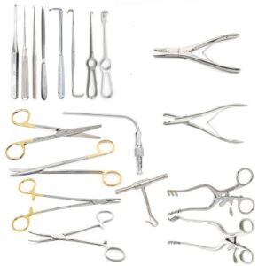
Where We Serve
Fizza Surgical exports to 50+ countries. Browse our country-specific pages with local regulatory guidance and pricing: