Bone Wax Application: Hemostasis in Orthopedic Surgery
Bone wax instruments and technique: how it works, correct dosing, risks, and absorbable alternatives for hemostasis in bone surgery.
 Made in Sialkot · Since 1980
The sternum is split, the retractor is in, and cancellous bone edges are weeping steadily from the marrow. Suction clears it; it comes straight back. This is the moment bone wax earns its place on the tray — a pea of soft wax smeared firmly into the bleeding cut surface, and the ooze stops mechanically in seconds. No coagulation cascade, no diathermy, just a physical seal pressed into the open trabeculae.
Bone wax is one of the oldest hemostatic aids still in daily use, and one of the most misapplied. Used correctly it is fast and cheap; used carelessly it interferes with the very bone healing the surgery is meant to restore. It is also one of the few consumables that lives on the tray rather than in the pharmacy — alongside the applicators and dissectors, bone wax instruments and the wax itself travel together to the field.
What Bone Wax Is and How It Works
Traditional bone wax is a sterile, non-absorbable compound — classically beeswax softened with a plasticiser such as paraffin or isopropyl palmitate. It carries no pharmacological action whatsoever. Its hemostasis is purely tamponade: worked into the cut cancellous surface, it plugs the open vascular channels and the marrow spaces, and bleeding stops because the blood physically cannot get out.
That mechanism is its strength and its limitation. Because it is inert and non-resorbable, it stays where you put it — which controls bleeding reliably but also means a permanent foreign body sits at the bone interface unless removed.
Where It Is Used
Bone wax appears anywhere a raw cancellous surface bleeds and pressure or cautery cannot reach it:
- Sternotomy — the divided sternal edges in cardiac surgery are the textbook indication.
- Craniotomy and neurosurgery — bleeding from the skull diploe and from bur holes, where cautery near the dura is undesirable.
- Orthopedic and spinal surgery — cut cancellous bone at osteotomy sites, iliac crest graft donor sites, and the raw surfaces exposed during arthroplasty.
- Maxillofacial and ENT — controlled oozing from facial bones and the mastoid.
Application Technique
Good technique is quick but not casual. The sequence that separates reliable hemostasis from a smeared, ineffective mess:
1. Prepare the Surface
Clear blood from the bleeding bone first. Wax adheres and seals far better against a surface that has been suctioned or briefly packed than against a flooded one. A dry-ish surface lets the wax key into the trabeculae rather than float on a film of blood.
2. Soften the Wax
Take a small amount — genuinely small, a pea or less for most edges — and warm it between gloved fingers until it reaches a pliable, putty-like consistency. Cold wax crumbles and will not seal; over-worked warm wax is easy to press home.
3. Press, Don’t Paint
Firmly smear and press the wax into the bleeding surface so it enters the open channels. The pressure is what creates the tamponade. A light surface coat that never engages the trabeculae will lift off with the next swab.
4. Use the Minimum Effective Amount
This is the rule that protects the patient. Apply only enough wax, for only the duration needed, to achieve hemostasis. Excess wax left in the wound is what drives the complications below. Where a bone surface must later heal or fuse — a spinal fusion bed, a fracture line — wax should be kept off the surfaces you want to unite.
The Risks Nobody Should Skip Over
Bone wax controls bleeding, but it is a permanent foreign body, and the literature is clear about what excess or careless use can cause:
- Impaired bone healing and fusion. Wax packed into a fracture or fusion site forms a physical barrier to osteoblast migration and new bone bridging. In spinal fusion this can contribute to non-union.
- Foreign-body and granulomatous reaction. The beeswax base can provoke a chronic granuloma — sometimes presenting months later as a mass or persistent inflammation.
- Increased infection risk. A retained inert bolus can act as a nidus for infection and has been shown experimentally to reduce the bone’s ability to clear bacteria.
- Migration and embolism. Rarely, a fragment can migrate or, in vascular fields, embolise.
None of these is a reason to abandon bone wax — they are reasons to use the smallest effective amount and to remove excess before closure, especially at sites intended to heal.
Bone Wax vs Absorbable Alternatives
The permanence problem has driven a class of water-soluble and resorbable hemostatic putties — alkylene-oxide (poloxamer-based) waxes and newer bioresorbable formulations that seal bleeding then dissolve or absorb over days, leaving no permanent barrier. They cost more, but where bone must fuse they remove the healing-impairment concern.
| Property | Traditional bone wax | Absorbable/water-soluble wax |
|---|---|---|
| Base material | Beeswax + plasticiser | Poloxamer / bioresorbable polymer |
| Hemostasis | Mechanical tamponade | Mechanical tamponade |
| Resorbed? | No — permanent | Yes — days to weeks |
| Effect on bone healing | Can impair union if excess | Minimal barrier once dissolved |
| Cost | Low | Higher |
| Best for | Sternum, skull, non-fusion sites | Fusion beds, graft donor sites |
Applicator Instruments
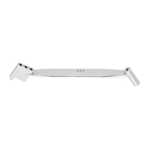
Most surgeons apply wax with a gloved finger or a Freer elevator, but dedicated bone-wax applicators exist — a shaped, blunt spatula or a small malleable delivery tip that presses wax precisely into a bur hole or a narrow osteotomy without contaminating surrounding tissue. In neurosurgery and deep spinal work, an applicator reaches surfaces a fingertip cannot. A Penfield or Freer dissector doubles as a serviceable applicator on most trays, which is why it usually lives alongside the practice’s other bone surgery instruments.
Removing Excess Before Closure
The discipline that matters most comes at the end of the case, not the beginning. Once hemostasis is achieved, surplus wax smeared beyond the bleeding surface should be lifted away — scraped off with a Freer or curette and irrigated — before the wound is closed. This is easy to skip when the field is dry and the surgeon has moved on, yet it is precisely the retained excess that seeds granuloma and infection weeks or months later.
At sites intended to unite — a spinal decorticated fusion bed, a fracture line, an osteotomy meant to knit — the rule is stricter still: keep the wax off the healing interfaces entirely, and where oozing there is a problem, favour an absorbable agent that will not sit as a permanent barrier between the bone ends. Reserve traditional wax for surfaces that do not need to fuse, such as the outer sternal table or the skull diploe.
Storage and Handling on the Tray
Bone wax is supplied as individually foil-wrapped sterile sticks, and it behaves differently with temperature. Straight from a cool store it is stiff and crumbly; a moment of hand-warming brings it to the pliable state that seals. Keep unopened wax at controlled room temperature, open it only when needed, and discard any opened remainder rather than returning it to the field. Because it is inert and carries no drug, it has a long shelf life — but the sterile barrier of the individual wrap is what keeps it safe to implant, so damaged packs are discarded.
Frequently Asked Questions
Is bone wax absorbed by the body?
Traditional beeswax-based bone wax is non-absorbable — it remains as a permanent foreign body where applied. Newer water-soluble and bioresorbable waxes are designed to dissolve or absorb over days, which is why they are preferred at sites that must heal or fuse.
Can bone wax stop arterial bleeding?
No. Bone wax controls bleeding from the raw cancellous surface of cut bone by tamponade. It does not seal a named vessel or arterial bleeder — those need ligation, a clip, or cautery.
Why is minimising the amount so important?
Excess wax is the root of nearly every reported complication — impaired bone union, granuloma, and raised infection risk. Applying only what is needed for hemostasis, and removing surplus before closure, keeps the benefit while limiting the foreign-body burden.
Should bone wax be used in spinal fusion?
With caution. It is fine on surfaces away from the intended fusion, but should be kept off the decorticated bone you want to unite, because it physically blocks bridging bone. Many surgeons prefer an absorbable hemostatic agent in the fusion bed itself.
In Summary
Bone wax remains a fast, dependable, inexpensive way to stop cancellous bone bleeding by mechanical seal — indispensable in sternotomy, craniotomy, and orthopedic work. The discipline is in the dose: prepare the surface, soften the wax, press it home, and use the least amount for the shortest time. Where bone must heal, reach for an absorbable alternative. Handled with that restraint, it is a hemostatic aid that has kept its place on the tray for over a century for good reason.
Need precision surgical instruments?
Configure complete instrument sets with our team — ISO 13485 certified, CE marked, made in Sialkot since 1980.
Related Guides
View all articles →
Where We Serve
Fizza Surgical exports to 50+ countries. Browse our country-specific pages with local regulatory guidance and pricing: